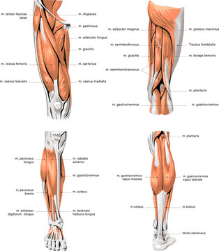
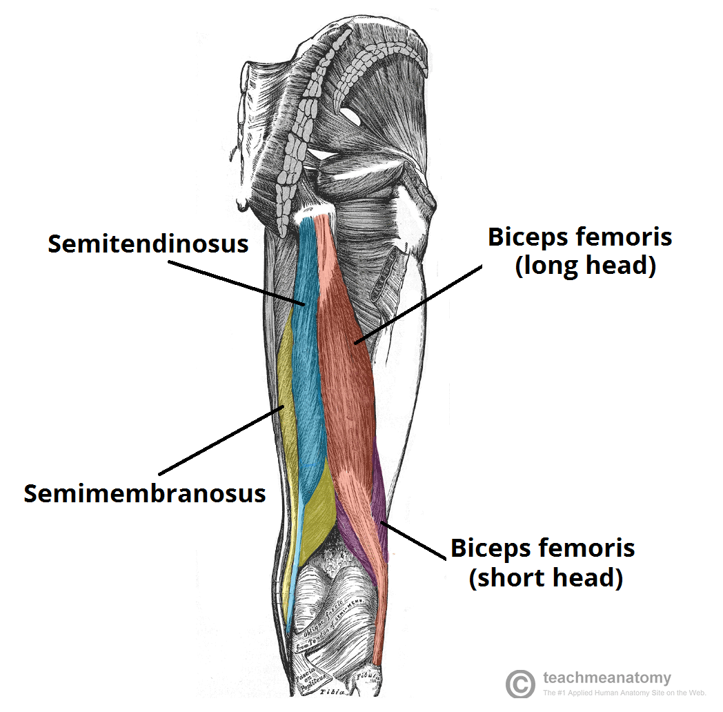
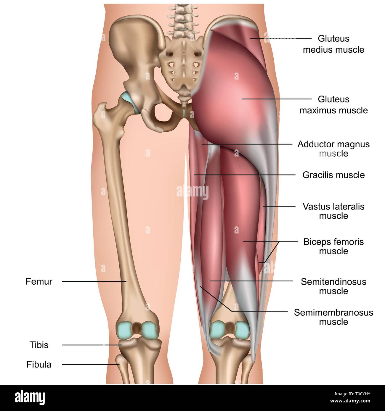
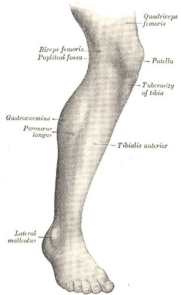
41 thigh muscle chart
Muscles of the Thigh - Anterior - Medial - TeachMeAnatomy Muscles of the Thigh; Muscles in the Anterior Compartment of the Thigh. View Article. Muscles in the Medial Compartment of the Thigh. View Article. Muscles in the Posterior Compartment of the Thigh. View Article. Anatomy Video Lectures. START NOW FOR FREE. TeachMe Anatomy. Part of the TeachMe Series. Leg Muscles: Thigh and Calf Muscles, and Causes of Pain - Healthline There are two main muscle groups in your upper leg. They include: Your quadriceps. This muscle group consists of four muscles in the front of your thigh which are among the strongest and largest...
3D muscle anatomy videos | Kenhub 04.08.2022 · Gluteus maximus muscle (3D) - hip flexion. The benefits of learning anatomy using 3D models has been debated at length over the past few years, with many studies proving that it’s not always what it’s been cracked up to be! That being said, there is one area of anatomy study where teachers and students alike agree that using 3D animations work best: learning muscle …
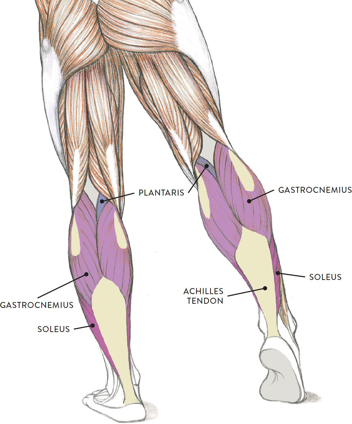
Thigh muscle chart
Leg Muscles Anatomy, Function & Diagram | Body Maps - Healthline Gastrocnemius (calf muscle): One of the large muscles of the leg, it connects to the heel. It flexes and extends the foot, ankle, and knee. Soleus: This muscle extends from the back of the knee to ... Quadriceps femoris muscle: Anatomy, innervation, function 22.07.2022 · Vastus medialis lies medial to rectus femoris and is partially covered by it. The sartorius muscle also crosses the superficial surface of vastus medialis. In the middle third of the thigh, vastus medialis forms the lateral wall of the adductor canal (Hunter’s canal).This canal is completed by adductor longus and adductor magnus posteriorly, and by sartorius medially. Causes and treatment of inner thigh pain - Medical News Today Mar 20, 2018 · Muscle injuries. The inner thigh muscles, or adductors, can become strained or torn by certain movements or activities. These can include running or turning too quickly.
Thigh muscle chart. Thigh muscles Diagram | Quizlet Start studying Thigh muscles. Learn vocabulary, terms, and more with flashcards, games, and other study tools. Lower Leg Anatomy, Diagram & Pictures | Body Maps - Healthline The main muscle in this area of the leg is the gastrocnemius, which gives the calf a bulging muscular appearance. Some nerves of the sacral plexus innervate this area, namely the superficial... Vaccine Administration: Needle Gauge and Length - Centers for … The vastus lateralis muscle in the anterolateral thigh can also be used. Most adolescents and adults will require a 1- to 1.5-inch (25–38 mm) needle to ensure intramuscular administration. Vaccines must reach the desired tissue to provide an optimal immune response and ; reduce the likelihood of injection-site reactions. Needle selection should be based on the: Route Age … Causes of Thigh Pain and When to See a Healthcare Provider Exercise has been proven to help thigh pain that involves your muscles, bones, ligaments, tendons, and nerves. This is known as your musculoskeletal system. If your pain is coming from your back, lumbar stretches and strengthening exercises may relieve pressure from spinal nerves. Exercises that correct your posture may also be helpful.
Muscles of the Hip - Anatomy Pictures and Information - Innerbody The adductor muscle group, also known as the groin muscles, is a group located on the medial side of the thigh. These muscles move the thigh toward the body's midline. Included in this group are the adductor longus, adductor brevis, adductor magnus, pectineus, and gracilis muscles. Hip and thigh: Bones, joints, muscles | Kenhub Function : Hip joint: Thigh extension, Thigh external rotation; Knee joint: Leg flexion, Leg external rotation. The medial compartment of the thigh is comprised of six muscles: gracilis, pectineus, adductor longus, adductor brevis, adductor magnus and obturator externus. Key facts about the medial thigh muscles. Leg Muscles: Anatomy and Function - Cleveland Clinic The muscles in your upper and lower legs work together to help you move, support your body's weight and allow you to have good posture. They enable you to do big movements, like running and jumping. They also help you with small movements, like wiggling your toes. Leg muscle strains are common, especially in the hamstrings, quads and groin. Muscles of the Anterior Thigh - Quadriceps - TeachMeAnatomy The muscles in the anterior compartment of the thigh are innervated by the femoral nerve (L2-L4), and as a general rule, act to extend the leg at the knee joint. There are three major muscles in the anterior thigh - the pectineus, sartorius and quadriceps femoris. In addition to these, the end of the iliopsoas muscle passes into the anterior ...
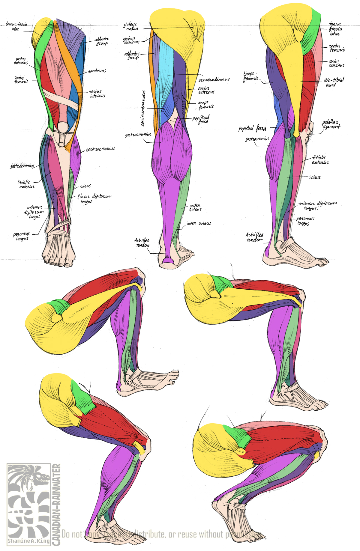
Muscles of the Leg - Chart for Massage School Notes calcaneus via achilles tendon. weak plantarflexion of the foot at ankle. may be absent in approx. 10% of people. Popliteus. lateral femoral condyle. posterior tibial surface above soleal line. laterally rotates femur, flexes the knee, unlocks knee from and extended position. deepest muscle of posterior knee. WebMD - Better information. Better health. The calf muscle, on the back of the lower leg, is actually made up of two muscles: The gastrocnemius is the larger calf muscle, forming the bulge visible beneath the skin. The gastrocnemius has ... Thigh Muscle Diagram | Leg muscles anatomy, Leg muscles ... - Pinterest Thigh Muscles Quad Muscles Anatomy Study Physical Fitness The Muscles of the Leg anatomy chart shows in every possible view the way that the muscles and other pieces of the leg work together in motion and flexibility. This chart is beautifully illustrated and offers the most comprehensive look at the muscles of the human leg available. Thigh Anatomy, Diagram & Pictures | Body Maps - Healthline Muscles in the medial thigh help to bring the thigh toward the midline of the body and rotate it. These muscles are the adductor longus , adductor brevis , adductor magnus , gracilis, and the...
Muscles of the Medial Thigh - TeachMeAnatomy The muscles in the medial compartment of the thigh are collectively known as the hip adductors. There are five muscles in this group; gracilis, obturator externus, adductor brevis, adductor longus and adductor magnus. All the medial thigh muscles are innervated by the obturator nerve, which arises from the lumbar plexus.
20 Sexy Thigh Tattoos for Women in 2022 - The Trend Spotter A thigh tattoo is only going to stretch noticeably with considerable muscle or weight gain. Skin is pretty elastic, so it’s going to take a lot to stretch your tattoo out of shape. If you take up weightlifting, your tattoo could change slightly with immense muscle growth, but for the most part, you probably won’t be aware. The most significant change you might see with weight gain …
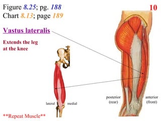
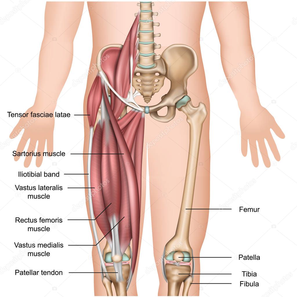
Thigh Muscles: Anatomy, Function & Common Conditions - Cleveland Clinic Quadriceps include four large muscles located in the front of the thigh: vastus lateralis, vastus medialis, vastus intermedius, and rectus femoris. They start at the pelvis (hip bone) and femur (thigh bone) and extend down to the patella (kneecap) and tibia (shin bone). Sartorius muscle is a long, thin muscle — the longest in the human body.
Femoral hernia: Symptoms, pictures, treatments, and more 15.11.2021 · A femoral hernia occurs when tissue pushes through the muscle wall of the groin or inner thigh. Learn when to see a doctor, what surgery entails, and more.
Muscle anatomy reference charts: Free PDF download | Kenhub Nov 03, 2021 · This muscle chart eBook covers the following regions: Inner hip & gluteal muscles; Anterior, medical and posterior thigh muscles; Anterior, lateral and posterior leg muscles; Dorsal and plantar foot muscles; This eBook contains high-quality illustrations and validated information about each muscle. It is available for free. Download free PDF (8 ...
muscle diagram leg muscles hip lower thigh diagram labeled kenhub limb anatomy femur extremity pelvis anterior posterior arm structures muscle joint upper bones. Deadlift Anatomy - YouTube . anatomy deadlift muscles animation motion 3d movement hinge leg press workout during training joints proper muscle human strength gluteus trainer.
Leg Muscles Diagram Pain Muscles Of The Leg - Part 1 - Posterior Compartment - Anatomy Tutorial . anatomy leg muscles posterior muscle diagram 3d anterior compartment lower human muscular body thigh drawing foot ankle knee spain. Leg muscles deep etc clipart left usf edu tiff medium. Leg lower shin anterior muscles labeled knee splints chandler physical ...
Muscles of the hips and thighs - Course Hero Figure 9-8. The superficial muscles of the thigh. Figure 9-9. The quadriceps group of four muscles. The view on the left has the rectus femoris cut away to show the vastus intermedius which is below it. The sartorius muscle is a distinctively long and thin muscle that crosses the thigh diagonally. It is visible in Figure 9-8.
Chart of Major Muscles on the Front of the Body with Labels - Health Pages A muscle of the medial thigh that originates on the pubis. It inserts onto the linea aspera of the femur. It adducts, flexes, and rotates the thigh medially. It is controlled by the obturator nerve. It pulls the leg toward the body's midline (i.e. adduction) Biceps brachii An upper arm muscle composed of 2 parts, a long head and a short head.
Muscle Charts of the Human Body — PT Direct For your reference value these charts show the major superficial and deep muscles of the human body. Superficial and deep anterior muscles of upper body Superficial and deep posterior muscles of upper body
Thigh - Wikipedia In human anatomy, the thigh is the area between the hip and the knee. Anatomically, it is part of the lower limb. The single bone in the thigh is called the femur. This bone is very thick and strong (due to the high proportion of bone tissue), and forms a ball and socket joint at the hip, and a modified hinge joint at the knee. Structure ...
Numbness in the thigh: Causes and treatment - Medical News … 16.09.2019 · Causes of numbness in the thigh include lupus, some autoimmune conditions, and multiple sclerosis. Treatment depends on the cause. Learn more here.
Anterior thigh muscles • Anatomy & Function - GetBodySmart The muscles of the anterior thigh include three members: sartorius, quadriceps femoris muscle and articularis genus muscles. The most notable member of the group is the quadriceps femoris, which consists of four heads: rectus femoris vastus medialis vastus lateralis vastus intermedius This powerful muscle crosses both the hip and knee joint, providing strong extension of the leg and flexion of ...
Average Thigh Circumference and Size in Males and Females Jul 02, 2022 · We'll explore the reasons for this in the upcoming thigh size chart section. After a man hits middle age, the average male thigh circumference decreases to 20.9 inches between the ages of 50 and 59 and 20.6 inches between the ages of 60 and 69. From there, the measurements decrease further still, reaching 19.5 inches between the ages of 70 and 79.
Upper Thigh Pain When Walking: Causes, Types and More Jun 20, 2022 · Thigh muscle soreness can be treated or cured in a variety of ways. If you experience thigh pain due to injury or overuse, you may start with an in-home exercise program. These types of home exercise programs usually involve stretches and strength training to help improve your overall strength and flexibility.
PDF MUSCLE ORIGIN, INSERTION, AND ACTION LIST CHARTS - Curofy Muscles that act at Hip (to move leg) ACTION ORIGIN INSERTION Pectineus Flexes hip, adducts leg Pubis and pubic ramus From lesser trochanter to superior part of linea ... 86 Muscle Origin, Insertion, and Action List Charts Muscles that Extend Knee (quadriceps work as a group, as when kick-ing a ball) ACTION ORIGIN INSERTION Vastus lateralis ...
Leg Muscle Anatomy, Function, & Diagrams - Study.com The thigh of the leg has three major muscle groups to move the leg forward, backward, and towards the midline of the body. These muscles surround and control the movement of the femur, which is the...
Healthhype.com Ž†&Ä¢'k9ä2¯ÉÞ G¢ RÞý K|J ˆª4H"/4ÚÞ´Ì %# ˜ C"45£Ø¶í#ÒðË7ÉS-«k&i¸Q:—Â8‹·O$‹ uP{ —ä ùñ ôÎ?îE#5'Fb¥ì ÓAERô-®ø!)JbjB…¤(u)‹ &cËJ ›idX κw wÒ Å—‹c0 ‚r 41^¦£Ú VÌÝ+¤\ÿc"c¢íC‚$ C#îYOyg
Ethically Made - Sweatshop Free | American Apparel Effortless basics and iconic fashion favorites for women, men and kids. The original basic, from tees to hoodies, denim and more.
Hip and thigh muscles: Anatomy and functions | Kenhub The gluteal muscles can be divided into two main groups: Large and superficial muscles which mainly abduct and extend the thigh at the hip joint. These are the gluteus maximus, gluteus medius, gluteus minimus, and tensor fasciae latae. Small and deep muscles which mainly externally rotate the thigh at the hip joint and stabilize the pelvis.
Leg Muscle Diagram Pictures, Images and Stock Photos Human thigh muscle anatomy for the education. Muscular system legs Human muscular system of legs in back view. Gluteus medius, gluteus maximus gastrocnemius and other muscles. Pelvis, leg and hip bones skeleton poster. Bodybuilding and strong body vector illustration Cardiovascular System of the Leg and Foot 19 century medical illutrsation.
hip muscle chart - anatomylesson101.z21.web.core.windows.net Hip anatomy thigh joint muscle posterior nerves muscles nerve supply kenhub diagram pelvic ligaments bones lumbosacral innervation knee. AIS Upper Body Workout Chart » Stretching USA we have 8 Pictures about AIS Upper Body Workout Chart » Stretching USA like #LowerBackPain in 2020 | Yoga anatomy, Hip flexor, Anatomy, Hip and thigh: Bones ...
Causes and treatment of inner thigh pain - Medical News Today Mar 20, 2018 · Muscle injuries. The inner thigh muscles, or adductors, can become strained or torn by certain movements or activities. These can include running or turning too quickly.
Quadriceps femoris muscle: Anatomy, innervation, function 22.07.2022 · Vastus medialis lies medial to rectus femoris and is partially covered by it. The sartorius muscle also crosses the superficial surface of vastus medialis. In the middle third of the thigh, vastus medialis forms the lateral wall of the adductor canal (Hunter’s canal).This canal is completed by adductor longus and adductor magnus posteriorly, and by sartorius medially.
Leg Muscles Anatomy, Function & Diagram | Body Maps - Healthline Gastrocnemius (calf muscle): One of the large muscles of the leg, it connects to the heel. It flexes and extends the foot, ankle, and knee. Soleus: This muscle extends from the back of the knee to ...


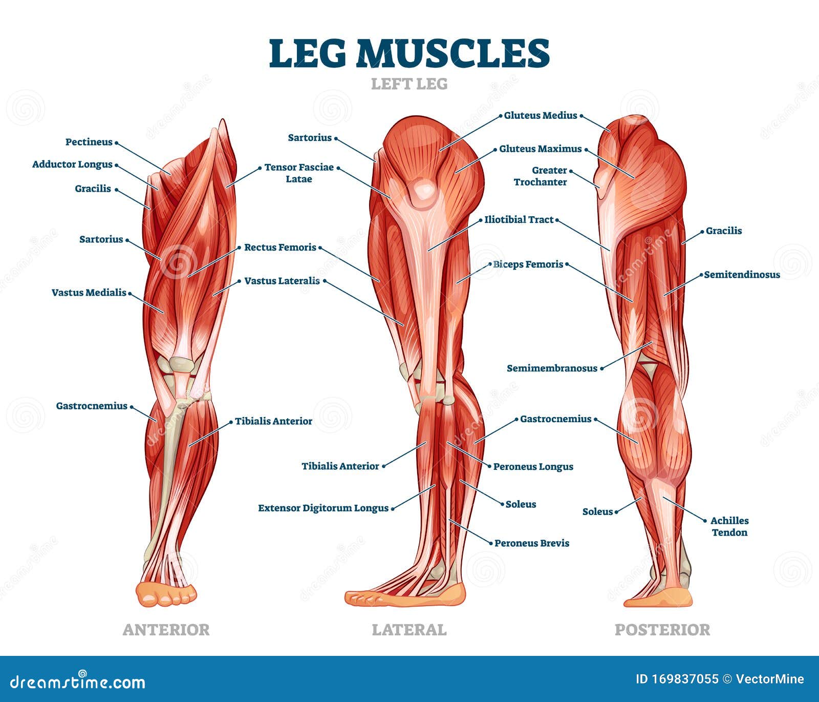

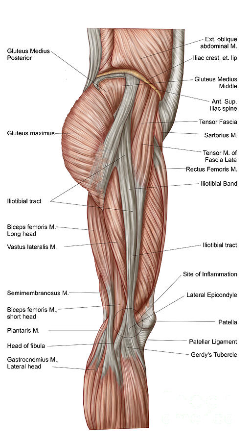

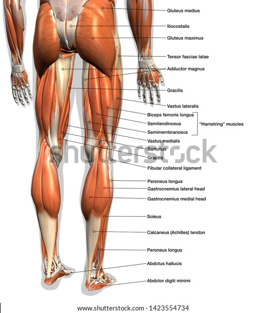






:background_color(FFFFFF):format(jpeg)/images/library/14013/Hamstring_muscles.png)





Post a Comment for "41 thigh muscle chart"